A body mapping computer system, FotoFinder, promises to offer the very latest in tracking and detecting potentially deadly cancerous moles, allowing patients to get care before there’s a problem. FotoFinder is a computerized mole mapping system that a doctor uses to create a detailed account of all the moles on a patient’s body. It uses a high-resolution camera to take the clearest possible photos, while sophisticated software compares skin marks to new ones each time a patient is scanned, detecting the tiniest of changes.
The goal is simple: To find problem moles or spots before they have a chance to turn into what could be deadly melanoma.
“A lot of our marks and moles aren’t anything to be worried about,” says Craig Pepple, vice president of n1health, a Richmond-based concierge medicine practice.
But for those that are worrisome, FotoFinder can find them. Then they can be treated.
Typically, a full body-scan in a dermatologist’s office involves a doctor going over a patient’s body by sight, noting spots and moles on the body. If an area looks suspicious, the doctor might remove it then and there for further testing. Or, the doctor might keep an eye on a mole, taking a digital photo for comparison and telling the patient to watch it for changes.
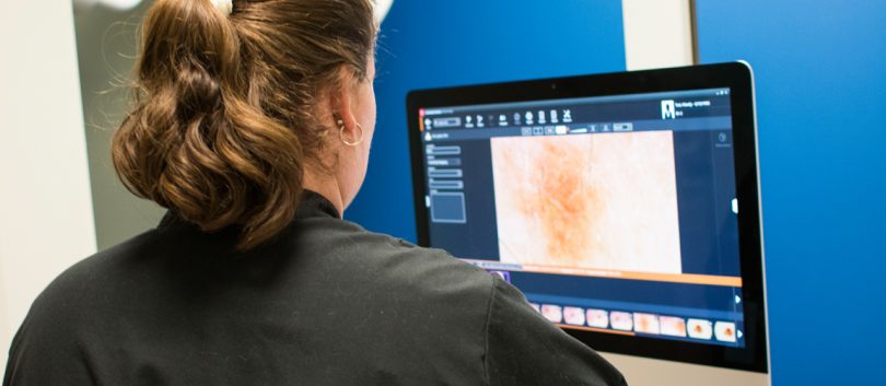
A scan by FotoFinder, meanwhile, involves taking numerous photos of the entire skin surface in just a few minutes, and then having the system quickly analyze the photos and determine the risk. The software identifies a mole three ways: Don’t worry, keep an eye on it, or do something now.
Brian Hamilton of Richmond had a scan done by a FotoFinder a few years ago when it picked up a suspicious mole on his ear. One particular mole had grown in size since a previous visit, and Hamilton was immediately referred to a specialist. A biopsy determined the mole was melanoma, and Hamilton ended up having the top part of his ear removed.
“Now I’m fine,” says Hamilton, who is in his late 40s, “although I often wonder what would have happened if I hadn’t been tested and my physician hadn’t found the problem when he did.”
Skin cancer is the most common cancer, with 1 in 5 Americans estimated to develop it in their lifetime, according to the American Academy of Dermatology. More than 8,500 people in the United States are diagnosed with skin cancer every day. About 75 percent of skin cancer deaths are from melanoma, a form of cancer that begins in cells that create the pigment melanin, and is one of the most dangerous cancer types leading to thousands of deaths per year.
If caught early, however, melanoma can be cut out before it has a chance to spread to other parts of the body. That’s why regular skin checks are highly recommended by the National Cancer Institute.
The company that makes FotoFinder was originally founded in Germany in 1991, later expanding to the United States. The FotoFinder bodystudio system was presented at the Congress of the German Dermatology Society in May 2013.
“Our skin is a large organ that takes a long time to examine,” Andreas Mayer, managing director of FotoFinder Systems in Bad Burnbach, Germany, told Business Wire in 2013. “Thanks to the impressive total body photographs, doctors can spot any conspicuous features and changes at a glance. In this way, the system supports the medical expertise of the dermatologist and gives patients more security.”
A FotoFinder machine isn’t cheap and isn’t covered by insurance, which is why it’s not available in most dermatology offices. The machine is available to members of n1Health, a health practice in which patients pay a yearly fee in order to have easy access to doctors, screenings with the latest technology and more intimate, specialized health care.
However, a FotoFinder body screening is also done during a one-time physical – no membership required — at the New Town center, including a follow-up visit to go over results.
A screening involves little more than standing in a few different poses against a blue screen while an automated camera attached to a robotic arm takes a series of photos. Afterward, the doctor provides the results on a CD, which can be given to a specialist if any follow-up is suggested.
“This technology is pretty amazing,” says Pepple. “It’s better than the human eye, and better than the human brain. It has the ability to catch something when it might be the difference between a little scar and a big scar.”
Hamilton says he probably would have noticed the mole on his ear eventually. But then, it might have been too late.
“Sometimes,” he says, “you just get lucky.”